Clinical Vignette 11
The 50th post on this blog! And perhaps it is fitting to put up this case, which I reviewed as a fairly new trainee in infectious diseases slightly more than a decade ago…
This young girl, who had just turned 18, developed upper respiratory tract symptoms and fever 4 days prior to hospitalisation. Her fever went up to 39 degrees Celsius 3 days later, and she started having streaks of blood in her (otherwise white) sputum. This was her chest X-ray when she presented to the emergency department (ED).

Chest X-ray of the 18-year-old girl with fever and hemoptysis.
Her white cell count was 1,590 per cubic millimetre of blood (the normal range is between 4,000 and 11,000 cells per cubic millimetre of blood), and she was also in acute renal failure. Because her oxygenation was exceedingly poor in the ED (SpO2 of 94% on 50% oxygen delivered via ventimask), she was transferred to the intensive care unit (ICU) for further monitoring, and started on broad-spectrum antibiotics – intravenous ceftazidime, intravenous azithromycin, and intravenous penicillin.
Within a day, she required inotropic support to maintain her blood pressure within physiological limits, and she required mechanical ventilation to support her breathing. A CT thorax showed extensive consolidation and cavitation within both lungs, worse on the right side.
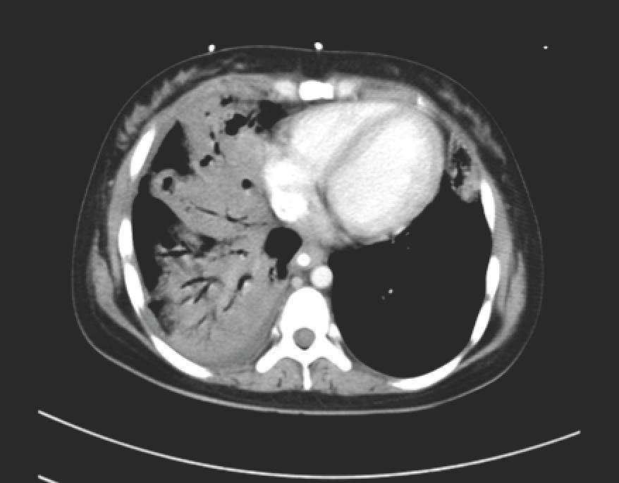
CT thorax showing predominantly right-sided consolidation and cavitation of the lungs.
Blood and sputum cultures were positive for bacteria.
Question: What bacteria can give rise to this clinical syndrome and what is the appropriate management for this patient?
[Updated 13th December 2014]
This clinical syndrome (young previously healthy patient with severe pneumonia especially after initial influenza-like symptoms, along with initial low neutrophil counts) is almost pathognomonic for necrotising pneumonia caused by Staphylococcus aureus strains that produce the Panton-Valentine leukocidin exotoxin (PVL). The only other syndrome (I know of) that approximates this is severe community-associated pneumonia caused by Streptococcus pneumoniae. PVL is a rather “diabolical” toxin that the organism extrudes into the immediate environment (hence exotoxin). It forms pores on the cell membranes of neutrophils, which causes these cells to lyse and release all their enzymes and free oxygen radicals, causing significant damage to the surrounding body tissue. In some ways, it can be considered a superantigen. Hence the lung destruction is not actually directly caused by the organism, but by the toxic stuff released from dying neutrophils.
The original case report can be found here in the Emerging Infectious Diseases journal archives. A case that actually resulted in my being hooked into staphylococcal research.

[…] Jerome Etienne at the end of 2003 – then director of INSERM E0230 – to discuss the case of PVL necrotising pneumonia in Singapore and to see if I could visit his laboratory. Much to my surprise, I was warmly welcomed (he […]
LikeLike