Clinical Vignette 59
A late middle-aged man with advanced lung cancer was switched to 3-weekly pembrolizumab after his malignancy progressed through multiple courses of chemotherapy. Pet CT at 12 weeks showed stable disease. However, he presented with sudden onset fever, worsening cough, and breathlessness after 6 months of immunotherapy. There was no contact or travel history of note, and clinical examination was remarkable only for mild hypoxia requiring oxygen support, fever of 38.7 degrees Celsius and bilateral basal pulmonary crackles. He did not have a “toxic” appearance. His chest X-Ray is shown below.
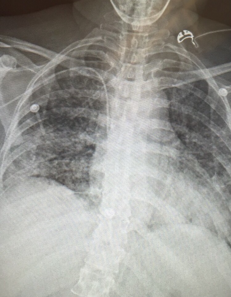
Question: What is the most likely diagnosis and how should this patient be managed?
[Updated 31 May 2016]
The diagnosis was correctly made by Dr. Eugene Lim (in the comment to this vignette), which is hypersensitivity pneumonitis secondary to pembrolizumab. This is a well known adverse effect of the class of cancer drugs known as checkpoint inhibitors. With regards to pembrolizumab, the incidence of pneumonitis is believed to be rare, occurring at approximately 3.8% of a large cohort of patients on the drug. Nonetheless, it is important to quickly recognise the syndrome as therapy is high-dose corticosteroids. There is a tendency in the local setting to view drug-related pneumonitis as diagnoses of exclusion, and to start patients on broad-spectrum antibiotics and/or antivirals first while arranging bronchoscopy and bronchoalveolar lavage – and to start steroids only after infection has been excluded. This delays appropriate treatment and subjects patients to unnecessary antimicrobial agents.

I’ll stick my neck out and call this autoimmune granulomatous alveolitis (or pulmonary alveolar proteinosis, or hypersensitivity pneumonitis, but I think I can see several large granulomas esp left UZ), due to uncontrolled T-helper/cytotoxic activation through PD-1L antagonism.
Treatment is with high dose steroid.
Of course the Resp/ID will balk at this and do bronchoscopic lavage and pan-cultures first. BAL should yield lymphocytes predominant PAS-positive proteinosis.
LikeLiked by 1 person