
Clinical Vignette 66
Not so much a clinical but a radiological vignette – a CT image from more than a decade ago.
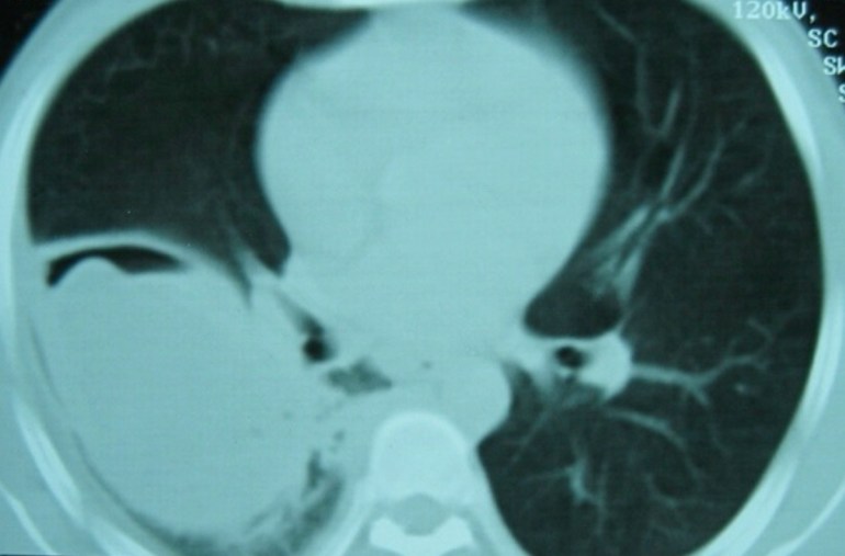
Question: What is the radiological sign and what is the diagnosis?
[Updated 20th September 2016]
The CT cut of the lungs shows a “water lily” sign (and thanks for those who suggested that it is an ugly or atypical “water lily”), which is almost diagnostic of a pulmonary hydatid cyst. This patient was seen more than a decade ago when I was in Peru for the famous Gorgas Diploma Course in Clinical Tropical Medicine. I cannot recommend this course highly enough, and there are now quite a number of distinguished alumni from Singapore.
Hydatid disease is caused by an infection by the canine tapeworm Echinococcus granulosus, and humans are an accidental intermediate host (the target intermediate hosts are sheep and cattle). It is still present in a large part of the world, including South America, New Zealand, Australia, Mediterranean countries, the Middle East, and parts of Asia. Hydatid disease most commonly manifests as liver cysts, although the tapeworm larva can also migrate to other organs, including the lungs. Patients are virtually always asymptomatic – the cysts typically grow at a slow rate of 1 cm per year. Treatment of symptomatic hydatid cysts is commonly by surgery (taking care not to rupture the cysts intra-operatively), or by puncture-aspiration-injection-re-aspiration (PAIR), where a scolicidal solution such as hypertonic saline or ethanol is injected. Anti-parasitic drugs such as albendazole work poorly.
