Clinical Vignette 35
A man in late middle-age, who in his youth had traveled extensively around the region, presented with nagging right upper abdominal discomfort for 2 weeks. He had no fever. The chest X-ray is shown below.
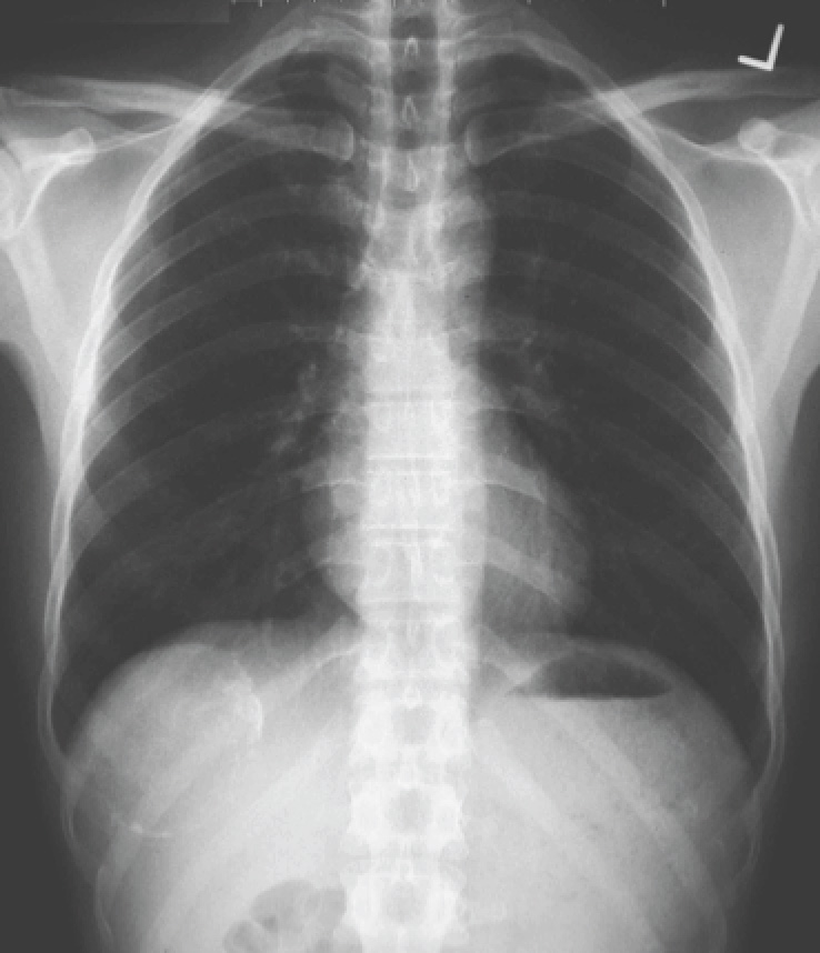
Chest X-ray of the man with right-sided abdominal pain.
Question: What is the abnormality seen and what infectious disease could this possibly be?
[Updated 6th June 2015]
There is a calcified cyst just below the right hemi-diaphragm. Given the location of the lesion, the question of an infectious disease, and the scope of the patient’s travels, it is likely to be a hydatid cyst. The corresponding CT image is shown below.
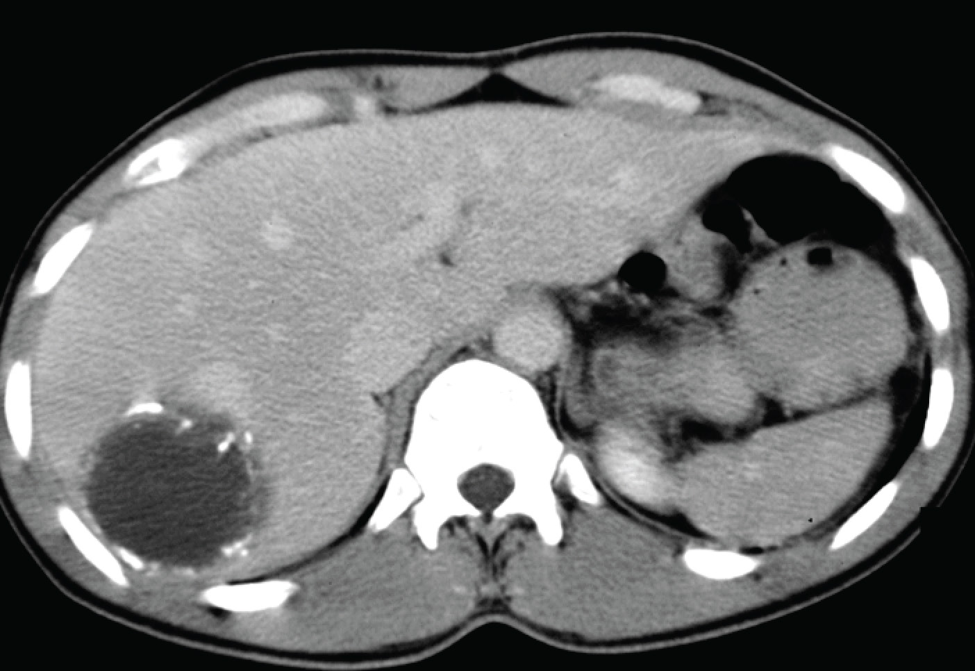
CT of the abdomen, showing a rounded cyst in the right lobe of the liver, with wall calcifications.
Hydatid cysts are the embryonal stages of the dog tapeworm, Echinococcus granulosus. The organism is present worldwide, particularly in sheep-farming areas of the world, although it is not endemic in Singapore or the U.S. Hydatid cysts are generally asymptomatic until they become too big (growing at approximately 1 cm per year), rupture (usually from trauma) or compress critical structures. When calcifications are seen, the hydatid cyst in question is dying (or dead). The cyst can be removed as a whole via surgery – it is important not to rupture the cyst during surgery otherwise an intense allergic/inflammatory response will occur with the spillage of cyst contents into the liver/peritoneum). Experienced radiologists may also perform PAIR (percutaneous aspiration, injection – usually of alcohol or some other sclerosant – and re-aspiration to collapse the cyst) as a less invasive but equally effective treatment of hydatid cysts. But PAIR is not routinely performed with calcified cysts.

X Ray showing calcified margin of a cyst within liver. It might be Hydatid cyst of liver.
LikeLike
That is excellent, Dr Akter!
LikeLike